Standing and walking become harder
Pain, heaviness or weakness in the legs may increase with walking or prolonged standing.

PROF. IAKIV FISHCHENKO · KYIV
Biportal endoscopic decompression, BESS/UBE, is surgery to relieve pressure on nerve structures in the lumbar spine.
The decision to operate depends on your symptoms, examination and complete MRI study. Not every case of stenosis needs surgery.
Send your MRI for a preliminary assessmentThe preliminary review is free. A response is provided on the day your scans are received.
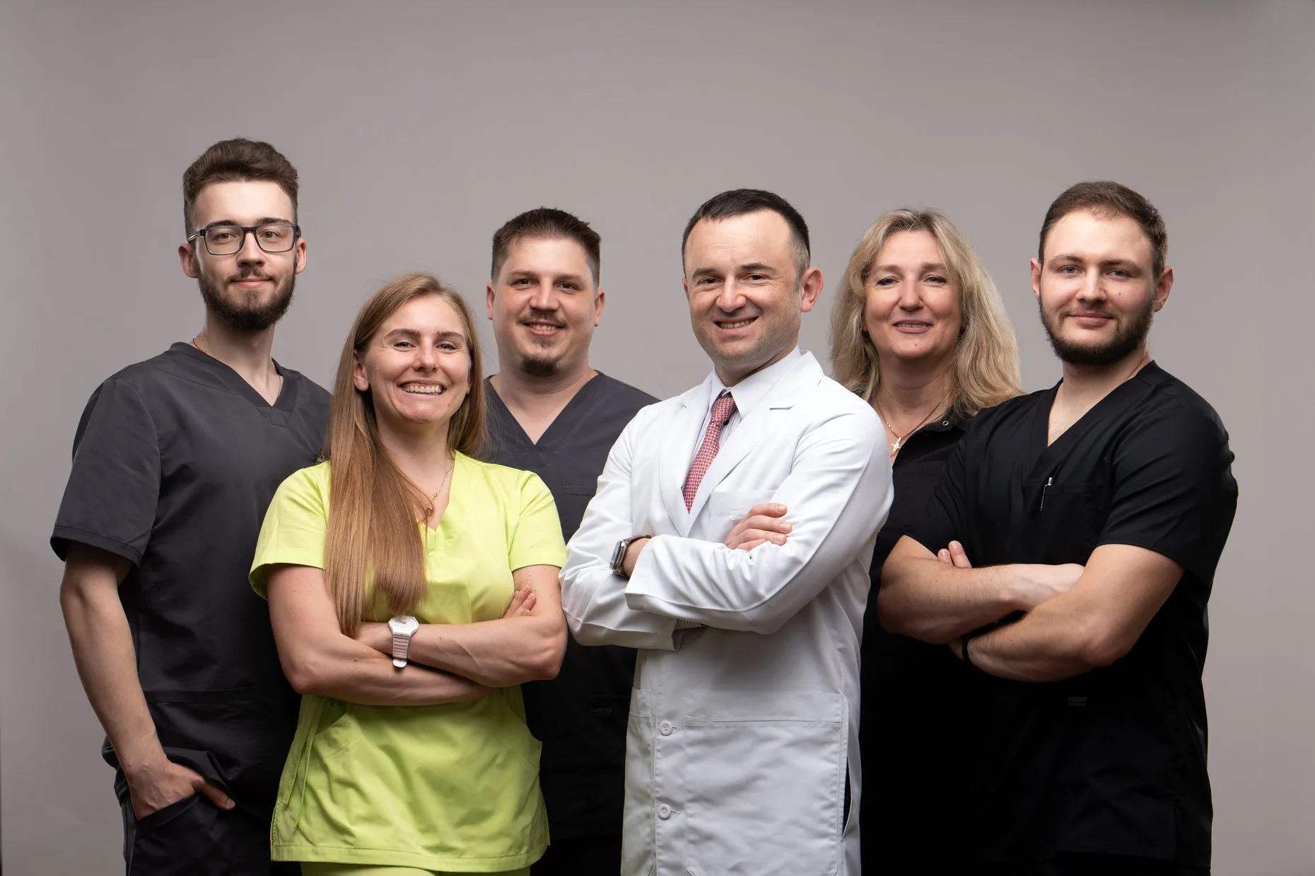
UNDERSTANDING YOUR DIAGNOSIS
Lumbar spinal stenosis is narrowing of the spinal canal or the spaces through which nerve roots pass. Joint changes, thickened ligaments and a bulging disc can contribute to this narrowing.
Pain, heaviness or weakness in the legs may increase with walking or prolonged standing.
Symptoms that ease when you sit down or lean forward are characteristic of neurogenic claudication.
Symptoms may affect one or both legs. Other conditions can cause similar symptoms.
Choosing treatment means matching the location and type of narrowing with your symptoms and neurological examination. If needed, the doctor may request CT or X-rays, including studies to assess spinal stability.
FROM DIAGNOSIS TO A DECISION
When your condition allows, treatment starts with tailored pain management, physical activity and rehabilitation. The approach depends on symptom severity.
Surgery is considered when symptoms substantially restrict daily life, treatment has not helped enough or neurological function is deteriorating.
Not everyone needs fixation. It is considered when there is instability, deformity or another individual indication.
COMPARING APPROACHES
Decompression aims to relieve pressure on nerve structures. The surgical approach and any need for fixation are chosen individually.
On a phone, swipe the table horizontally.
| What we compare | BESS / UBE | Microsurgical decompression | Decompression with stabilisation |
|---|---|---|---|
| How the surgeon works | The endoscope and instruments are introduced through two separate access points. | The surgical field is viewed through a microscope via a surgical approach, which may be a minimally invasive tubular approach. | Nerve decompression is combined with fixation and, when indicated, spinal fusion. |
| Main purpose | Remove tissue that compresses the nerves while taking the segment's stability into account. | Relieve pressure on the nerves under microscopic guidance. | Relieve nerve compression and address instability or deformity. |
| When it is considered | When the anatomy of the stenosis, symptoms and surgical plan allow endoscopic decompression. | When this approach suits the anatomy and the aims of decompression. | For specific indications; a diagnosis of stenosis alone does not mean implants are needed. |
| Implants | Usually not used for decompression alone. | Usually not used for decompression alone. | Fixation devices are used according to the surgical plan. |
| Recovery | Depends on the extent of surgery, your condition before treatment and your individual rehabilitation plan. | Depends on the extent of surgery, your condition before treatment and your individual rehabilitation plan. | Fixation and the process of bone fusion also need to be taken into account. |
| What to bear in mind | Endoscopy does not remove every risk or guarantee that an already damaged nerve will recover. | Microsurgery remains one of the options for decompression. | Fixation increases the extent of surgery and needs a clear indication. |
BESS/UBE offers small, separate access points, magnified imaging and targeted instrument control to help preserve surrounding tissue. Some studies have reported lower pain medication requirements and shorter hospital stays. Clinical outcomes can nevertheless be comparable to microsurgery; the benefits and pace of recovery depend on the individual case. When indicated, endoscopic decompression can be combined with stabilisation.
BESS / UBE IN ACTION
BESS and UBE refer to a biportal endoscopic technique. A camera is introduced through one access point, while instruments are used through the other under visual guidance.
The camera and instruments work through separate small access points.
The surgeon removes parts of the altered tissues causing compression.
The extent of nerve decompression is checked under endoscopic visual guidance.
A CONVERSATION WITH THE PROFESSOR
A video interview with Professor Iakiv Fishchenko about lumbar spinal stenosis. Watch the explanation and prepare your questions for a consultation.
PREPARATION AND RECOVERY
Provide your complete MRI/CT study and medical records. Tell the team about your regular medicines and other health conditions. Agree the required tests, food, drink and medication instructions with the anaesthetist.
The patient gets up after recovering from anaesthesia and having their condition assessed. The first time is supervised by a rehabilitation specialist. Discharge depends on the patient's condition and the extent of surgery.
Walking, exercises, any need for a brace, and returning to work and sport are agreed with the rehabilitation specialist. There is no single recovery timetable for every patient.
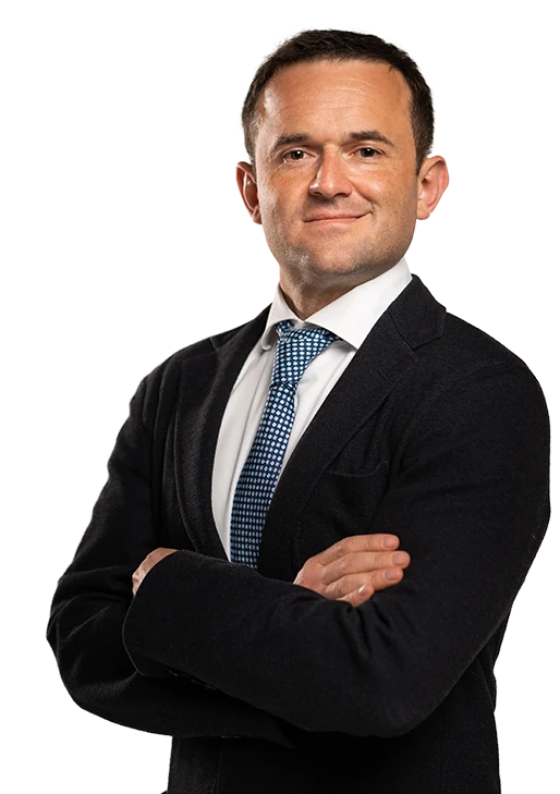
YOUR SURGEON
Doctor of Medical Sciences, professor and physician of the highest qualification category. Specialties: orthopaedics and traumatology, and neurosurgery. Practising in the specialty since 2004.
Author of an atlas of endoscopic spine surgery.
President of the Ukrainian Association of Endoscopic Spine Surgery. Full member of the Scoliosis Research Society, AO Spine and the American Academy of Orthopaedic Surgeons.
Honoured Doctor of Ukraine. Recipient of the President of Ukraine Prize (2008), a diploma from the Presidium of the National Academy of Medical Sciences of Ukraine (2013) and the Vesalius Award of the American Association of Neurological Surgeons (2013).
Professional profilePATIENT LIBRARY
The professor's explanations, BESS materials and answers to questions.
Open the resourceGeneral information, treatment, preparation, rehabilitation and contacts.
Open the resourceIllustrated instructions: a folder named with the patient's full name in Ukrainian, a ZIP archive, Google Drive and WeTransfer.
Open the resource →COST
The cost of endoscopic decompression depends on the extent of surgery and whether additional stabilisation is needed. Ask the administrator what the treatment includes: consumables, anaesthesia, hospital stay and follow-up appointments.
Administrator: +38 094 821 08 30 ↗No. MRI findings are assessed alongside symptoms and an examination. Nonsurgical treatment may be appropriate if there are no urgent indications. Surgery is considered when daily life is substantially restricted, treatment has not helped enough or neurological function is worsening.
Stenosis means narrowing of the space for nerve structures. A herniated disc can be one cause; changes in joints and ligaments often also contribute. The decompression plan therefore depends on all the causes of compression.
Biportal endoscopic decompression is one surgical option for lumbar stenosis. Its suitability is determined after assessing the anatomy, affected levels, stability and the patient's condition.
Not always. If decompression without stabilisation is sufficient, fixation implants are usually unnecessary. Instability, deformity and other factors may change the surgical plan. When indicated, endoscopy-assisted fusion is possible: an interbody implant (cage) is placed under endoscopic guidance, while screw fixation is performed percutaneously. The suitability of this approach and the type of implants are determined individually.
Not necessarily. Recovery of nerve function depends on the severity and duration of the damage. Some symptoms can persist even after adequate decompression.
You first get up after recovering from anaesthesia and having your condition assessed, under the supervision of a rehabilitation specialist. Further activity and return to work are planned individually.
Your complete MRI/CT study in DICOM format: all the contents of the disc in one ZIP archive, or a download link. Name both the folder and the archive using the patient's surname, first name and patronymic in Ukrainian. Include the radiology report, a description of symptoms and the patient's full name in the message.
The preliminary review is free. A response is provided on the day your scans are received.
Send your complete MRI/CT study, briefly describe your symptoms and provide the patient's surname, first name and patronymic. If the archive is large, send a link to it.
This information helps you prepare for a consultation. An individual treatment plan is developed after an examination and review of your scans.